

In fertility treatment, embryos can now be genetically tested even before transfer. Until now, however, this has required the removal of cells from the embryo. A new method aims to eliminate this step entirely: Instead of biopsying the embryo, the DNA it releases into the culture medium during its development is analyzed. New research shows how far this technique has come—and where its limitations lie.
The Embryo’s DNA Ends Up in the Culture Medium
Artificial insemination does not automatically result in the formation of an embryo. In the laboratory, the embryos continue to develop for a few days before doctors and embryologists decide which embryo should be transferred to the uterus. Various pieces of information are available during this process. The embryo’s appearance and development can be assessed, and in certain treatments, genetic testing may also be considered. So-called preimplantation genetic diagnosis—or preimplantation genetic testing—can be used, among other things, to examine chromosomal abnormalities. The problem: For traditional testing, genetic material must be obtained directly from the embryo. This is precisely where the so-called non-invasive preimplantation genetic testing comes into play.
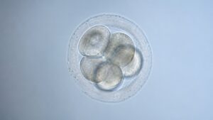
During IVF, embryos develop in a special culture medium. Researchers have found that so-called cell-free DNA can enter the culture medium during this development. This DNA contains genetic information that may originate from the embryo. If the fluid is analyzed after the embryo has developed, it may be possible to determine the embryo’s chromosomal makeup. The major difference from the traditional method is that the embryo itself would not need to be punctured or biopsied to obtain this DNA.
The procedure is referred to as non-invasive PGT-A, or niPGT-A for short. The “A” stands for testing for aneuploidy, i.e., an abnormal number of chromosomes. This concept has been the subject of research for several years. Earlier studies showed that DNA can be extracted from the culture medium. At the same time, the results varied significantly from one study to another. A review published in 2023 therefore concluded that the clinical significance of the method had not yet been sufficiently established.
Why a Test Without a Biopsy Would be Interesting
In conventional PGT-A, a small number of cells are removed from the outer cell layer of the embryo at the blastocyst stage. This so-called trophectoderm biopsy then provides the material for genetic analysis. The method is well-established, but it involves an invasive procedure on the embryo.
A non-invasive test could therefore offer several advantages. No direct intervention on the embryo would be necessary, and the genetic testing could potentially be more easily integrated into the IVF process. There is also another scientifically interesting aspect: The traditional biopsy examines only a small area of the embryo’s cells. However, embryos can contain cell groups with different genetic compositions. This so-called mosaicism can complicate the interpretation of genetic test results. This is another reason why researchers hope that analyzing the released DNA could provide additional information.
A Large Study Provides Important New Data
Just how well the method actually works in embryo selection was recently investigated in a large randomized study published in September 2026 in Nature Communications. Women aged 35 to 42 participated in the study. The study was conducted at 13 fertility clinics in China. A total of 1,189 participants were randomized; 1,152 patients were included in the analysis. The study compared embryo selection using niPGT-A with conventional selection based on morphological criteria—that is, based on the appearance and development of the embryos.
The most important finding: The pregnancy continuation rate was 38.7 percent in the niPGT-A group and 36.8 percent in the group where embryos were selected based on their morphology. This difference was not statistically significant. No statistically significant difference was observed in the live birth rate either. It was 37.0 percent in the niPGT-A group and 35.5 percent in the comparison group.
In contrast, a difference was observed in early miscarriages: they occurred less frequently in the niPGT-A group. Overall, however, the authors point out that the study does not provide a sufficient basis for the routine use of niPGT-A in this patient group.
Technical Accuracy Also Remains a Challenge
The fact that embryos release DNA into the culture medium does not automatically mean that this DNA provides a perfect representation of the entire embryo. A large multicenter study from 2026 analyzed the culture medium from 2,539 blastocysts for this purpose. The researchers compared the genetic results from the released DNA with those from conventional trophectoderm biopsy.
While such studies demonstrate the method’s potential, they also make it clear that the concordance is not perfect. Another study from 2025 found an overall concordance rate of 63.6 percent between the analysis of DNA from the culture medium and conventional PGT-A in 100 samples examined. The authors therefore concluded that, at that time, the method was not yet reliable enough to replace the traditional test.
Possible Sources of Error
A key issue is the small amount of DNA. The culture medium contains significantly less genetic material than a direct cell sample. This can make it more difficult to generate a reliable genetic profile. Added to this is the risk of contamination. Not all DNA found in the culture medium necessarily originates from the embryo. DNA from other cells or other contaminants can also influence the analysis.

A recent review from 2026 therefore identifies several key challenges: a sometimes low amount of DNA, potential maternal or other DNA contamination, dealing with embryonic mosaicism, and a lack of uniform laboratory standards. This makes it clear why developing a non-invasive test is more difficult than it initially sounds.
Non-Invasive Testing Does Not Automatically Mean Better Results
For patients, one question is particularly crucial: Does the new test ultimately lead to a higher probability of a successful pregnancy and live birth? This is precisely where the new randomized study provides important insight. Although the method can extract genetic information from the culture medium, the study group showed no statistically significant advantage in terms of continued pregnancy or live birth.
This is a key distinction between a technically interesting test and a clinically proven improvement in fertility treatment. A procedure can reliably detect genetic differences yet still fail to demonstrate any additional benefit for treatment. For use in reproductive medicine, therefore, it is not enough for the laboratory results to be accurate. Ultimately, what matters is whether the procedure actually improves treatment outcomes.
Research is Already Moving on to the Next Step
Scientists are no longer merely investigating whether DNA can be extracted from the culture medium at all. They are also trying to determine which characteristics of this DNA are particularly informative. For example, a study published in September 2026 analyzed the so-called fragmentation patterns of cell-free DNA from the culture medium. Among other things, fragment lengths, end structures, and other molecular characteristics were examined and compared with the chromosomal status of the embryos.
Such approaches could help extract more information from a very small amount of DNA in the future. Artificial intelligence and so-called multi-omics methods are also being discussed as possible next steps in development. This would allow various types of biological information to be combined, rather than relying exclusively on a single genetic marker.
What the Method Could Mean for Future IVF Treatments
If niPGT-A proves reliable in further studies, it could change embryo selection in IVF in the long term. Instead of extracting cells directly from the embryo, it might be possible to analyze a portion of the culture medium. The embryo itself would remain untouched. Until then, however, further clinical studies are necessary. In particular, it must be clarified how reliable the genetic results are under different laboratory conditions and whether the method actually leads to better treatment outcomes.
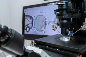
Research to date therefore paints a mixed picture: The technical foundation is in place, but its clinical application has not yet been conclusively established. The large randomized study from 2026 is an important step in this regard because it examines not only the technical accuracy of the procedure but also its actual impact on pregnancy outcomes. However, it was unable to demonstrate a significant advantage over conventional morphological embryo selection.
A Possible Future – But Not Yet a Replacement for Biopsy
The idea is fascinating: An embryo could be genetically tested without having to extract cells from it. The DNA required for this may already be present in the fluid in which the embryo develops in the IVF laboratory. However, there is still an important research step to be taken between this biological possibility and a reliable clinical application.
The current state of the art can therefore best be summarized as follows: Non-invasive genetic embryo testing is among the most interesting developments in modern reproductive medicine. It could one day offer an alternative to biopsy, but the data available so far are not yet sufficient to routinely replace the conventional method. Further randomized studies and standardized laboratory procedures will need to determine whether this promising technology can indeed become a new standard method for embryo selection.
You may also be interested in...
-

New research may afford future predictability of IVF success, as an abnormal gene expression within…
-
You are likely wondering what comes next after having successfully completed your IVF process and…
-
A medical process known as Preimplantation Genetic Diagnosis (PGD) may help would be parents determine…