A groundbreaking IVF technique developed in Newcastle to reduce the risk of mitochondrial disease has resulted in the birth of eight babies, according to a published study. All eight babies show no signs of mitochondrial DNA disease. The babies, four girls and four boys, including a pair of identical twins, were born to seven women who were at high risk of passing on a serious disease caused by mutations in mitochondrial DNA. The findings, published by the Newcastle team that pioneered mitochondrial donation using fertilized human eggs, suggest that the new treatment, known as pronuclear transfer, effectively reduces the risk of otherwise incurable mitochondrial DNA diseases.
Mitochondrial DNA Mutations: A Hereditary Disease that can be Devastating
The results were published in two articles in the New England Journal of Medicine (NEJM) and describe the reproductive and clinical outcomes of the pronuclear transfer treatments performed to date. All babies were healthy at birth, achieved their developmental milestones, and the disease-causing mitochondrial DNA mutations from the mother were either undetectable or present in concentrations that are very unlikely to cause disease. The technique was developed on human eggs by a team from Newcastle University, UK, and Newcastle upon Tyne Hospitals NHS Foundation Trust in work funded by Wellcome and NHS England.

The mother of a girl born after mitochondrial donation said: “As parents, we wanted nothing more than to give our child a healthy start in life. IVF with mitochondrial donation made this possible. After years of uncertainty, this treatment gave us hope – and then it gave us our baby. When we look at her now, full of life and possibilities, we are overwhelmed with gratitude. Science has given us a chance.“ The mother of a little boy added: ”We are now the proud parents of a healthy baby boy – a real success story for mitochondrial replacement therapy. This breakthrough has dispelled the heavy cloud of fear that once hung over us.
The NHS Mitochondrial Reproductive Care Pathway offers women with mitochondrial disorders a range of reproductive options, including mitochondrial donation as part of a research study. Professor Sir Doug Turnbull of Newcastle University, a member of the Newcastle team, said: “Mitochondrial disorders can have a devastating impact on families. Today’s news gives many more women at risk of passing on this condition new hope that their children can grow up without this terrible disease. Within the NHS and in a well-regulated environment, we can offer mitochondrial donation to affected women in the UK as part of a research study.”
Every year, around one in 5,000 children are born with mitochondrial DNA mutations that can lead to serious diseases. Mitochondria produce the energy necessary for life and contain a small piece of DNA that contains only part of the information needed for energy production. Harmful mutations in mitochondrial DNA can lead to reduced energy availability, which particularly affects tissues with high energy requirements, such as the heart, muscles, and brain. Mitochondrial DNA is inherited from the mother, so these diseases are passed from mother to child. Men can be affected but do not pass on the disease. Despite years of research, there is still no cure for people with mitochondrial DNA disorders.
Pronuclear Transfer
Since there is no cure for mitochondrial DNA disorders, research is focusing on IVF-based technologies to reduce the risk of disease by limiting the transmission of disease-causing mitochondrial DNA mutations from mother to child. The new IVF-based mitochondrial donation technology, pronuclear transfer, which was legalized in the UK in 2015, aims to reduce the risk of mitochondrial diseases in children born to women with a high proportion of disease-causing mutations in their mitochondrial DNA. The Newcastle team has now included pronuclear transfer in a research study, along with a range of reproductive options available to women at risk of passing mitochondrial diseases to their children.
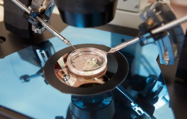
The technique, known as pronuclear transfer, is performed after fertilization of the egg. It involves transplanting the nuclear genome (which contains all the genes essential for our individual characteristics, such as hair color and height) from an egg carrying a mitochondrial DNA mutation into an egg donated by an unaffected woman from which the nuclear genome has been removed. The resulting embryo inherits the nuclear DNA of its parents, but the mitochondrial DNA is predominantly inherited from the donated egg cell.
The Study on Reproductive Outcomes
The British team from Newcastle that developed and optimized pronuclear transfer for use in fertilized human eggs is now reporting the results of pronuclear transfer treatment to reduce the risk of mitochondrial DNA diseases. The proportion of disease-causing mitochondrial DNA detected in babies after pronuclear transfer treatment ranged from undetectable to 16% in the blood of newborns. The presence of mitochondrial DNA mutations in babies born after pronuclear transfer treatment is due to the transfer of maternal mitochondria surrounding the nuclear DNA at the time of transfer. The transfer of maternal mitochondrial DNA is a known limitation of mitochondrial donation technologies.The team is seeking to better understand and resolve this issue as part of a fundamental research program.
Professor Mary Herbert, lead author of the study on reproductive outcomes conducted by Newcastle University, said: “The results give cause for optimism. However, further research to better understand the limitations of mitochondrial donation technologies is essential to further improve treatment outcomes. “Mitochondrial donation procedures are currently considered risk-reducing treatments due to the transfer of maternal mitochondrial DNA during the mitochondrial donation process. Our ongoing research aims to bridge this gap between risk reduction and prevention of mitochondrial DNA diseases by addressing this issue.”
Pronuclear transfer treatment is offered as part of an integrated program that also includes preimplantation genetic diagnosis (PGD) to reduce the risk of mitochondrial DNA disorders. In accordance with HFEA regulations, pronuclear transfer is only offered to women who are unlikely to benefit from PGT treatment. At the time of reporting on the integrated program of PGT and pronuclear transfer, clinical pregnancies were confirmed in 8 of 22 (36%) patients who underwent pronuclear transfer and in 16 of 39 (41%) patients who underwent PGT. Pronuclear transfer resulted in eight births and one additional pregnancy. PGT resulted in 18 births. In children from pronuclear transfer, the concentrations of disease-causing mitochondrial DNA mutations were either undetectable or well below the levels at which disease symptoms are observed.
The Publication on the Clinical Results
The Newcastle team describes the treatment pathway developed to provide the best possible care for women with pathogenic mitochondrial DNA mutations. It details how the mothers of the first children born using this technique were monitored and supported during pregnancy and how their babies were closely observed from birth. Some of the mothers already had symptoms of mitochondrial disease, including vision loss and heart problems. Others had family members with the condition and remain at risk of developing symptoms and passing them on.

All eight babies, including a pair of identical twins, were healthy at birth and are developing normally according to the description—five of them have had no health problems since then. In the publication, the team points out that three babies have overcome some early health problems that it believes are not directly related to the mitochondrial donation. The team in Newcastle offers counseling and treatment to women with harmful mitochondrial DNA mutations in the UK. They are carefully monitored during pregnancy and after mitochondrial donation. Six out of seven women had no complications. One woman experienced a rare pregnancy complication with high blood fat levels (hyperlipidemia), which responded well to a low-fat diet.
All eight babies, including the twins, were born by normal vaginal delivery or planned cesarean section. All babies had a normal weight for their gestational age. The level of disease-causing mitochondrial DNA mutation was measured in blood and urine cells and was undetectable in five babies. Three babies had low levels of disease-causing mitochondrial DNA mutations—5% and 9%, 12% and 13%, and 16% and 20% in blood and urine, respectively. These levels are well below the 80% required for clinical disease. The researchers note that at the 18-month follow-up, the disease-causing mutation was not detectable in the child with 5% and 9% in the blood and urine. All children are participating in an 18-month developmental study, and at the time of reporting, all babies had reached the appropriate developmental milestones.
One child developed transient startle responses (with neck flexion and eye blinking) at 7 months of age, which resolved after 3 months without treatment. Another child who was breastfed developed high blood lipid levels (hyperlipidemia), which also affected the mother during pregnancy, and was successfully treated with a low-fat diet. This child was also diagnosed with an abnormal heart rhythm (arrhythmia), which is being successfully treated with a reduced dose of antiarrhythmic drugs. (Although children born after PGT are not routinely followed up, the team notes that one child was found to have a heart abnormality.) A third child had a urinary tract infection, which responded quickly to antibiotic treatment. The authors say that the children’s health conditions are not thought to be related to the mitochondrial DNA mutations in their mothers, as the low levels found in these babies are unlikely to cause any symptoms of disease. Symptoms of these mutations only occur at concentrations above 80%. It would be expected that the pronuclear transfer procedure itself would manifest clinically in a more uniform manner, i.e., affecting all children in the same way. However, follow-up studies are of utmost importance to identify patterns in childhood diseases.
Longer-Term Follow-Up of Children is Essential
The team emphasizes that follow-up studies are essential for identifying patterns of childhood disease and states that it will continue the studies until the children reach the age of 5. Professor Bobby McFarland, Director of the NHS Highly Specialised Service for Rare Mitochondrial Disorders (Newcastle Hospitals NHS Foundation Trust) and Professor of Pediatric Mitochondrial Diseases at Newcastle University, is the lead author of one of the publications.
He said: “Although longer-term follow-up of children born after mitochondrial donation is of paramount importance, these initial results are very encouraging. It is a great privilege to see the joy and relief these children have brought to their parents.” The Lily Foundation, a charity dedicated to fighting mitochondrial diseases, has supported the work in Newcastle. “They too are delighted with the results of these published studies, as for many affected families this is the first real hope of breaking the cycle of this hereditary disease.
You may also be interested in...
-

There is a potential correlation between factors that affect a woman's fertility and her risk…
-
Pelvic inflammatory disease, or PID, affects more than 1 million women in the United States…
-

Conditions that affect the heart are broadly called heart disease. Conditions that involve blocked or…