
Researchers at the Francis Crick Institute have developed a new stem cell model of the mature human amniotic sac that replicates the development of the tissues that support the embryo two to four weeks after fertilization. This is the first model of amniotic sac development after two weeks. As described in a study published in Cell, the new model can be used to study the formation and function of the human amnion and help identify previously unknown ways in which the amniotic sac may support embryonic development. It also shows promise for medical procedures that use the amniotic membrane.
Replication of the Amniotic Sac
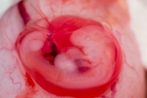
The amnion is a protective membrane that expands into a sac filled with amniotic fluid, which leaves the body when the amniotic sac ruptures before birth. Until recently, it was believed that its main function was to surround and protect the baby, cushion it from shocks, and allow nutrients to pass through before the placenta forms. Researchers have not been able to study the amnion in detail because current stem cell embryo models do not typically represent the later stages of human development, and human embryos cannot be studied beyond 14 days for ethical reasons.
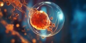
The team at the Crick developed a new 3D model – called the post-gastrulation amnioid (PGA) – that closely resembles the human amnion and other supporting tissues after gastrulation (when the cells of the embryo organize themselves into layers that form tissues and organs). To do this, they cultivated human embryonic stem cells in a series of steps using only two chemical signals over a period of 48 hours, after which the cells organized themselves into the inner and outer layers of the amnion. By the tenth day, a sac-like structure had formed in over 90% of the PGAs. This gradually enlarged over a period of 90 days without any further signals being given. The cell composition in the models showed remarkable similarity to the human amniotic sac, and the fluid in the PGAs mimicked the contents of human amniotic fluid.
Interaction Between the Amnion and the Embryo
Using genetic manipulation techniques, the researchers found that a transcription factor (a gene that switches other genes on or off) called GATA3 impaired the growth of amnion tissue when it was switched off in PGAs. Conversely, when GATA3 was enhanced in embryonic stem cells, the cells developed an amnion-like structure without receiving any further signals. These experiments showed that GATA3 is necessary for the initiation of amnion development.
Finally, the team investigated whether the amnion tissue not only protects the embryonic cells but also supports their development and specification. They mixed PGA cells with untreated embryonic stem cells and found that the untreated cells formed amnion-like structures, suggesting that signals from the amnion may indeed communicate with the embryonic cells. Due to its regenerative, anti-inflammatory, and antimicrobial properties, the amniotic membrane from women who have opted for a cesarean section can be donated and used for medical procedures such as corneal reconstruction, uterine lining repair, or the treatment of burns and ulcers.
Since transplanted tissue relies on donations, the team believes that PGAs could be an alternative source of amniotic membranes that could even be grown from a patient’s own cells. The researchers are now working with the Crick’s translation team to investigate the potential of PGAs in the clinic and to better understand the communication between the amniotic membrane and embryonic cells.
A New Perspective on the Amnion
Silvia Santos, group leader of the Quantitative Stem Cell Biology Laboratory at the Crick and senior author, said: “Early human development is still a black box due to ethical and technical limitations, but our new model gives us insights into this critical phase without the need to use human embryos. This work changes our view of the amnion as a purely protective structure: it is actively communicating with the embryo and promoting its growth. We are also excited about the potential of PGAs as a fast, inexpensive, and scalable way to provide amniotic membranes for medical purposes.”
Borzo Gharibi, senior research scientist at the Quantitative Stem Cell Biology Laboratory at the Crick and first author, said: “We originally investigated how embryonic cells form their identity, and then our research took a very interesting turn that led to the development of PGAs. This model replicates the mature human amniotic membrane for the first time and mimics the structural and functional characteristics of the human amniotic sac with high reproducibility. This provides researchers with a tool to study later stages of human development and the functions of the tissues that support the embryo.” This project was made possible through close collaboration with the genomics, metabolomics, and proteomics teams at the Crick.
You may also be interested in...
-

Polycystic ovary syndrome (PCOS) is a common endocrine disorder that affects millions of women worldwide,…
-
Most human eggs never have the chance to mature into eggs, a recent study sheds…
-
Researchers at the Center for Genomic Regulation (CRG) in Barcelona have identified a potential new…