
Research conducted by doctoral student Bohan Chen in the laboratory of Idse Heemskerk at the Department of Cell and Developmental Biology at the University of Michigan Medical School and his colleagues is improving a popular experimental model, thereby providing more insight into the internal processes during a critical phase of embryonic development. Scientists are studying development in order to understand, among other things, what can go wrong during the formation of body structures. The ultimate goal is to prevent birth defects and identify the causes of miscarriages.
New Insights Into Embryonic Development
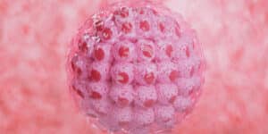
However, studying embryos in the laboratory raises important ethical and technical considerations. To address these concerns, many researchers use simple 2D structures made from stem cells called gastruloids. Gastruloids are grown or cultivated in vitro (in a Petri dish) and model some of the earliest moments of development without the possibility of developing into a human being. In experiments, gastruloids replicate aspects of a process called gastrulation.

During gastrulation, a primitive streak of cells develops into the three embryonic layers that ultimately form the body plan: the ectoderm (outer layer), from which the skin, nervous system, and other external structures develop; the mesoderm (middle layer), from which the heart, muscles, bones, and other internal structures develop; and the endoderm (inner layer), from which the gastrointestinal tract, lungs, liver, and other organs develop. However, in vitro cultivation of gastruloids could usually only be maintained for up to two days, after which the cells became disorganized and did not develop further. “We experimented with different media and made several other improvements to the model,” said Heemskerk.
Expanded Model to Answer More Questions
When the researchers then tried to cultivate the cells for longer than two days, it actually worked and some really interesting things happened. First, the team noticed that the stem cells of the developing mesoderm began to move beneath the original single cell layer and form a multilayered structure, just like in a real embryo. “This process is very difficult to visualize; we don’t even know how it works in mice,” said Heemskerk. However, thanks to their expanded model, they were able to see that the mesoderm stem cells migrate from the edge of the cell group to the center. This means that there is something – they don’t yet know what – that tells them which direction to move in.
The experts now have a constellation in which they can determine what controls their movement. This is crucial for finding out what goes wrong in certain cases, such as the development of a congenital heart defect. The team also found that mesoderm cells occur in several different subtypes in a gastruloid, expressing different genes, which they made visible using fluorescence.
By looking at which gene the cell expresses, it is possible to determine, to a certain extent, which organ it will ultimately develop into. According to Heemskerk, this raises the question of whether cells already know their fate before migration, or whether their final destination determines what they will become. Heemskerk hopes to continue this work with her expanded model to answer these and other questions about mammalian development. “This is a simple model that allows us to see things that would be very difficult to detect in a complex 3D structure, but it also captures biological phenomena and at the same time spares us the problems that arise when working with embryos.”
You may also be interested in...
-

A new approach by scientists at Kyoto University, published in Cell Genomics, promises a breakthrough…
-
A new discovery by researchers at the RIKEN Center for Biosystems Dynamics Research (BDR) in…
-
When a couple’s wish to have children isn’t coming true, male infertility is responsible one…