
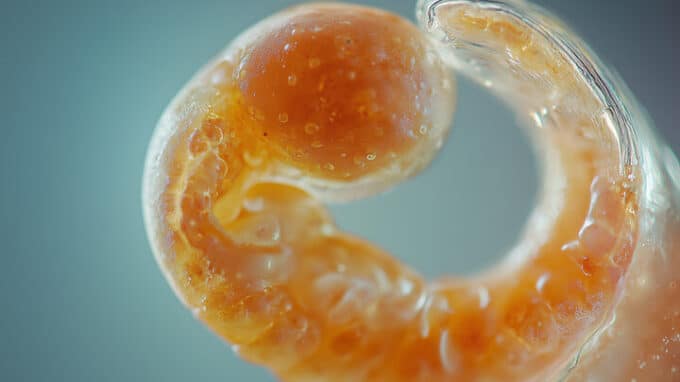
Researchers at the University of Adelaide have used cameras designed for quantum measurements to image embryos for the first time. Scientists at the university’s Centre of Light for Life investigated how ultra-sensitive camera technology, including the latest generation of cameras capable of counting individual packets of light energy at each pixel, can best be used in the life sciences.
Advances in Clinical IVF
The center’s director, Professor Kishan Dholakia, explained that the sensitive detection of these packets of light energy, known as photons, is crucial for capturing biological processes in their natural state, as it allows researchers to illuminate living cells with gentle doses of light. “Damage from illumination is a real problem that is often overlooked. Using the lowest possible light intensity in conjunction with these highly sensitive cameras is important for understanding the biology of living and developing cells,” said Professor Dholakia. He added that modern imaging technology is very exciting because it provides researchers with new insights.

The research team, which also included Zane Peterkovic, Dr. Avinash Upadhya, Ramses Bautista Gonzalez, Dr. Megan Lim, Dr. Chris Perrella, Admir Bajraktarevic, and Associate Professor Kylie Dunning, who also heads the Reproductive Success Group at the Robinson Research Institute, tested the embryo imaging technology in a preclinical study and published its findings in APL:Photonics. These samples are living, developing specimens that serve as the basis for studies supporting advances in clinical IVF. Digital camera technology has advanced to the point where fundamental physical concepts such as quantum mechanics become important and relevant, said lead author and doctoral student Peterkovic. Many natural compounds in cells glow when illuminated, which can tell researchers a lot about what they are seeing, but unfortunately the signal is very weak.
A large part of the project involved developing a method to fairly compare the image quality of different cameras. The analysis of the images was made possible by a combination of expertise in optics, biology, laser physics, and microscopy. The researchers even investigated how AI can be used to remove noise from the captured images, which is essentially static because the camera has difficulty capturing enough light. These steps go beyond simply placing a camera on a microscope to capture images. Future approaches to this work include expanding into the field of quantum imaging, where quantum states of light can be used to gain further information about the sample.
You may also be interested in...
-

Although some studies suggest that artificial insemination may have adverse effects on future offspring, there…
-
Understanding the IVF timeline could play a crucial role in a couple’s conceiving a child.…
-
Researchers have discovered that germ cells, which develop into eggs and sperm, cause sex-dependent differences…